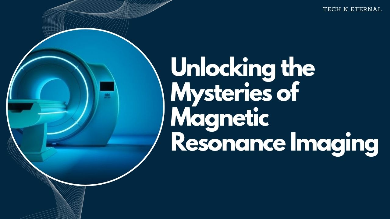
Magnetic Resonance Imaging (MRI)
Magnetic resonance Imaging (MRI) is a non-invasive medical imaging technique used to visualize internal structures of the body in detail. MRI leverages the principles of nuclear magnetic resonance to create highly detailed images of tissues, organs, and other structures inside the body, making it an invaluable tool in modern medicine.

How MRI Works?
The basic principle behind MRI (Magnetic Resonance Imaging) involves the use of strong magnetic fields and radio waves to excite hydrogen atoms within the body. Since hydrogen atoms are abundant in water and fat, which constitute a large portion of human tissues, they serve as excellent targets for MRI.
- Magnetic Field: The patient is placed within a powerful magnetic field, typically ranging from 1.5 to 3 Tesla. This magnetic field aligns the magnetic moments of the hydrogen nuclei.
- Radiofrequency Pulse: A radiofrequency pulse is then applied, which perturbs the alignment of the hydrogen nuclei. When this pulse is turned off, the nuclei relax back to their original alignment, releasing energy in the process.
- Signal Detection: The released energy is detected by the MRI (Magnetic Resonance Imaging) scanner and converted into digital signals. These signals are then processed by a computer to create detailed images of the internal structures.
Types of MRI
There are several types of MRI, each tailored for specific applications:
- Functional MRI (fMRI): Used to measure and map brain activity by detecting changes in blood flow.
- Cardiac MRI: Focuses on the heart and blood vessels, providing detailed images of cardiac structures and function.
- Musculoskeletal MRI: Used to visualize muscle, tendons, ligaments, and bones.
- Diffusion MRI: Measures the diffusion of water molecules in tissue, often used in brain imaging to access conditions such as stroke.
MRI Procedure

- Preparation: Before the scan, patient may need to remove metallic objects and wear a hospital gown. If contrast agents are required, an intravenous (IV) line is inserted to administer the contrast.
- Scanning: The patient lies on a motorized table, which slides into the MRI scanner. It is important to remain still during the scan to ensure clear images. The procedure is painless, but the scanner can be noisy, so earplugs or headphones are often provided.
- Duration: An MRI (Magnetic Resonance Imaging) scan typically takes between 30 to 60 minutes, depending on the complexity and area being examined.
Applications of MRI
MRI (Magnetic Resonance Imaging) is used across a wide range of medical fields due to it’s ability to provide high resolution images without exposing to ionizing radiation. Some common applications include:
- Neurology: Diagnosis brain tumors, multiple sclerosis, and neurological disorders.
- Orthopedics: Assessing joint abnormalities, spinal conditions, and soft tissue injuries.
- Cardiology: Evaluating heart conditions, blood flow, and vessel abnormalities.
- Oncology: Detecting and staging cancers, monitoring treatment response, and guiding biopsies.
- Gastroenterology: Visualizing abdominal; organs, liver disease, and inflammatory bowel conditions.
Advantages of MRI
- Non-Invasive: MRI does not involve ionizing radiation, making it safer for repeated use.
- High Contrast Resolution: Excellent for differentiating between different types of soft tissues.
- Multi-planar Imaging: Images can be obtained in any plane, providing comprehensive views of structures.
Limitations of MRI

- Cost: MRI scans can be expensive due to the advanced technology and equipment required.
- Time: MRI scans take longer compared to other imaging modalities like X-rays or CT scans.
- Contraindications: Not suitable for patients with certain implants, such as pacemakers or cochlear implants, due to the strong magnetic field.
Safety Considerations
- Metallic Implants: Patients with metal implants need to inform their healthcare provider before undergoing an MRI.
- Claustrophobia: Some patients may experience anxiety or discomfort due to the enclosed nature of the MRI scanner. Open MRI machines are an alternative but may have lower resolution.
- Contrast Agents: Gadolinium-based contrast agents are generally safe but can cause allergic reactions in rare cases. Patients with kidney problems should discuss risks with their doctor.
In summary, MRI is a versatile and powerful imaging technique that provides detailed and high quality images of the body’s internal structures. It plays a crucial role in the diagnosis., treatment planning, and monitoring of various medical conditions across different specialties.